
 Open Access
 Abstract
Objective: Neuroblastoma (NB) is the most common cause of solid cancer-related high death in children. Previous research shows that tumor microenvironment that involved Collagen that plays an important as heterogeneous role in its tumor microenvironment remodeling. However, less study has been described the collagen expression in the NB. Therefore, this study aimed to elucidate the expression of two prominent collagens (COL11A1 and COL4A1) which can be express abnormally in the NB cancer cohort in Vietnam.
Method: We collect the plasma circulating NB tumor cells and biopsies from City Children Hospital HCM city. These samples were operated with droplet digital RT-PCR method to evaluate the mRNA expression of two collagen subtypes: COL11A1 and COL4A1 on five patient biopsies. Internal control is run with GAPDH. Characterisation of clinicopathological traits (such as: age, gender, histology, MYCN amplification and stages) is correlated with the relationship of COL11A1 and COL4A1 mRNA levels.
Results: The results largely confirmed the optimization of COL11A1 and COL4A1 primers and qualified products in droplet digital RT-PCR with internal GAPDH control. In addition, there is a higher COL11A1 mRNA expression in NB than in healthy control. Moreover, higher COL11A1 was significantly linear with high-risk classification (such as stage 3 or 4 comparing versus stage 1 or 2; MYCN amplification versus MYCN no amplification) in this cancer. In contrast, many of the associations between NB and COL4A1 were non-linear. Other clinicopathological traits (such as: age, gender, histology) did not show any significant differences for COL11A1 and COL4A1.
Conclusion: Altogether, this study confirms the COL11A1 could be a potential biomarker to evaluate the prognosis of NB disease. The integration of proteomic and genomic profiling should be added with larger cohort to address the value of COL11A1 in the future.
INTRODUCTION
NB is a pediatric tumor originating from neural crest cells 1 . It is a heterogeneous characteristic and may contribute by epigenetic regulation during neural differentiation. Despite multiple cancer-related recommendations for treatment, diagnosis, and follow-up, strengthening cancer patients' long-term predictions remains low survival rate 2 . In particular, more than 60% of effective therapy can be operated in developed nations, but only 30% of successful therapy is recorded in developing countries, including Vietnam 3 . Therefore, it is crucial to find appropriate markers in cancer diagnosis and prognosis.
There are currently at least 28 different collagen proteins that divided into four subtypes based on their super-molecule collection, including fibril-forming collagens, three disrupted helical chains of collagen-binding to fibril, network-forming collagens, and membrane anchor collagens 4 . Type XI collagen is extracellular minor fibrillary collagen, which codes for the α1 chain of pro-collagen 11A1 and mature COL11A1 5 . COL11A1 levels have also been found to be elevated in many tumor forms, including colorectal, pancreatic, breast, and non-small-cell lung cancer, relative to normal tissues, which can be a target marker for the differential diagnosis of invasive breast carcinoma. COL4A1 encodes the chain of COL4A1 and secretes it into an extracellular matrix 6 . Overregulated COL4A1 facilitates tumor invasion in bladder cancer cells via tumor budding activation 6 . Overexpressed COL4A1 also leads to breast cancer cell proliferation and migration 7 .
To evaluate the two collagens in NB, we examine the different expression levels of COL11A1 and COL4A1 received on NB liquid biopsy. The study focus on assessing the expression of COL11A1 and COL4A1 comparing between NB and healthy donors.
MATERIALS AND METHODS
Sample collection and circulating tumor cell isolation from BP
All samples taken from those diagnosed with NB at City Children’s Hospital at Ho Chi Minh City. Ethic reviewer’s committee asserts the ethical document at the Ho Chi Minh City Children’s Hospital. The peripheral blood sample of healthy control (BL01) was used as a positive control for experimental design of primers ( Table 1 ). Four peripheral blood (PB) samples were NB05, NB06, NB07, NB08 and one tissue sample (NB09) from NB patient. Circulating tumor cells were collected as previously report. In brief, whole PB shipped in 10 mL blood collection tubes (BCT) containing a cell-free RNA preservative. Collect up to 4 mL of plasma from centrifuged and Isolate circulating RNA using a commercially available kit Qiagen that captures exosomes, platelets and cell-free RNA from plasma. Clinical information such as the patient's age, stage of the disease, and MYCN amplification status was provided by the hospitals.
Total RNA extraction
The total RNA will be extracted by using the ThermoFish Company’s GeneJET RNA purification kit. After extraction, total RNA was quantified and qualified using the nanodrop (Thermo Scientific) and gel electrophoresis, respectively. The GeneRuler 100 bp DNA Ladder was used. For tissue, a quantity of around 30 mg per sample was grinded with 1 ml of RMPI. The whole mixture was centrifuged to remove supernatant before 300 μl of Lysis Buffer (20 μl of 14,3 M β-mercaptoethanol was applied to each 1 ml amount of Lysis Buffer) and 600 µl of diluted Proteinase K were added. They were centrifuged for 10 minutes at 12000 g after incubated at room temperature, and then the supernatant was transferred into a new tube. 450 μl of ethanol (96-100 %) was applied. 700 μl of lysate was moved into the purification column inserted in a collection tube. Then, they were centrifuged at 12000 g for 1 minute. The flow-through was discarded and the column of purification was inserted back into the collection tube. This process was replicated before all the lysates in the column were transferred. In the purification column, 700 μl of Wash Buffer 1 and 600 μl of Wash Buffer 2 was added and centrifuged for 1 minute at 12000 g, respectively. Then, the flow-through was discarded and the column of purification was put back into the collection tube. After, 250 μl of Wash Buffer 2 was added and centrifuged for 2 minutes at 12000 g. The collection tube was discarded, and a sterile 1,5 mL RNase-free micro-centrifuge tube was transferred to the purification column. Finally, 50 μl of nuclear-free water was added to the membrane of the purification column. They were centrifuged at 12000 g to elute RNA for 1 minute. The column was discarded and the purified RNA was used.
For the blood sample, 0.5ml of whole blood was centrifuged at 14000 rpm for 10 minutes. The plasma was removed after centrifugation and the pellet was re-suspended in 600 μl of Lysis Buffer, adding 20 μl of 14.3 M β-Mercaptoethanol each 1 ml Lysis Buffer amount. 450 μl of ethanol (96-100 %) was applied. The remaining steps were performed in the same way as tissue sample.
Semi quantitative RT-PCR
Primer design
Primers are designed using an Origene website (OriGene Technologies Inc.). OriGene offers a battery of products for gene expression. The specific primers are checked using Primer blast to perform specificity checking and avoid primer self-complementary.
First-strand cDNA synthesis
The procedure was performed according to the manufacturer's instructions using the Revertaid 1st cDNA Synth Kit of Thermo Scientific. The total RNA was synthesized to cDNA. The total reaction volume was 20 μl, including primer set 10 pmol, extracted RNA, 5X Reaction Buffer, RiboLock RNase Inhibitor (20 U/µL), 10 mM dNTP Mix, RevertAid RT and distilled water. They were mixed gently and centrifuged briefly. Then, they were incubated at 42°C, 60 minutes. The reaction was completed by heating at 70°C for 5 minutes.
Digital droplet RT-PCR
The cDNA was diluted (1:1000) ratio and amplified using the Thermo Scientific DreamTaq Green PCR Master Mix kit on the digital droplet PCR-Qiagen. RT-PCR was set up 2 minutes at 95 for initial denaturation and 30 cycles, each consisting of 30 seconds at 95, 30 seconds at 55 , and 1 minute at 72. The close cycle for 10 minutes at 72. GADPH was used as a control. The PCR ingredients for 1 reaction were 21 µL of water, nuclease-free, 2 µL of diluted cDNA, 1 µL of each primer and 25 µL of DreamTaq™ Green PCR Master Mix. The total volume of the reaction was 50 μl reaction. For droplet generation, transfer 20 µL PCR mix to sample wells on the droplet generation cartridge. Add 70 µL of droplet generation oil and add droplet generation and then transfer droplets to PCR plate. Aspirate and dispense the droplets slowly, over 5 to 6 s each, without touching the opening of the tip to the droplet cartridge.
Amplification products were analyzed by gel electrophoresis. The Gene-Ruler 1 kb Plus DNA Ladder was used. 4 μl of products were separated on 2% gel-containing agarose gel in a 0.5X TBE buffer at 100V in 30 minutes. The PCR results were obtained by using UV Trans-illuminator.
Data analysis
Data visualization and correlation were performed with Prism 8 (Graph-Pad Software Inc). The expression of the phenotype was performed by the t-test. Values expressed as means ± SD and P value <0.05 are statistically significant.
RESULTS
Quantitative and qualitative results of the total RNA collected from samples
The total RNA was quantitated by using the nanodrop (Thermo Scientific). Finally, total RNA products were checked by using gel electrophoresis.
The concentration was the highest in the NB09 specimen (132.16 ng/μl). The concentration of blood samples, by comparison, was lower. Four samples of blood under 60 ng/μl were obtained, the lowest total RNA concentration was recorded in sample NB07. The 260/280 ratio (approximately 2) indicates pure RNA ( Table 2 ).
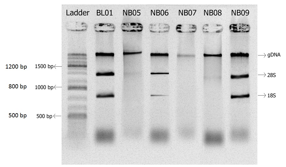
All gDNA bands appear in the above of well. We can see 2 clear bands of 28S and 18S rRNA in lanes of BL01, NB06 and NB09 samples. In lanes of NB05 and NB08, there was no presence of the 18s band. In lane NB07, there were no both 18S and 28S of rRNA ( Figure 1 ). The theoretical 28S:18S ratio was around 2:1, and this ratio suggests the intact RNA. However, further research was required to see whether there was a relationship between the amount of RNA profiles and mRNA integrity.
The optimal GAPDH within blood and tissue samples
Optimization of the GAPDH is important for internal control and is quantitative to COL11A1 and COL4A1 level. The product of mRNA was shown the best at 55°C. which illustrated in Figure 2 .
Figure 2 . The GAPDH amplification at control RNA and BL01 samples.

*A: The cDNA was diluted (1:1000); B: The cDNA was diluted (2:1000); C: The cDNA was diluted (3:1000)

Both BL01 and control had the expression of GAPDH at approriate band of 532 bp. Particularly, the BL01 was much lower than that of the healthy control ( Figure 3 ). Moreover, the results in Figure 4 showed that GAPDH expressed in both blood and tissue samples. The length of the products was 532 bp.
Optimization of amplification products of COL4A1 and COL11A1 with blood and NB samples
Figure 4 . Blood and NB samples are examined by two different primer sets towards COL4A1(A) or COL11A1(B) expressions

The gel electrophoresis result from Figure 4 showed the successful PCR product amplification reaction. Specifically, the appearance of products at 120bp and 140bp, respectively. There was no discernible change in most samples in most samples in COL4A1 expression, except where no expression was observed in the tissue sample. However, the COL11A1 gene showed a significant difference between the patients diagnosed with NB compared with the normal control.
Testing the expression of the COL11A1 and COL4A1 on NB samples
Figure 5 . Violin box plot of mRNA levels that normalized to GAPDH. (A). Comparison between COL11A1 and COL4A1; (B). Comparison between stages and MYCN amplification status
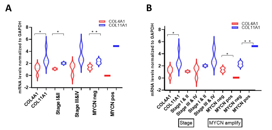
The mRNA levels normalized to GAPDH of COL4A1 and COL11A1 were significant differences (P<0.05) between each group ( Figure 5 A). Comparing with the stage I-II, there was a significant difference in the expression of COL4A1 and COL11A1 (P<0.05). MYCN non-amplification status has a significant difference between COL4A1 and COL11A1 (P<0.01). No significant difference was found in Stage III-IV and MYCN amplification ( Figure 5 A). There were no significant differences in the expression of COL4A1 and COL11A1 in the early stage compared to the later stage. The opposite is true for the MYCN amplification ( Figure 5 B).
Correlation of collagen expression with NB clinic pathological parameter
COL11A1 expressed positive in all samples of patients diagnosed with NB at both early (stage 2) and later stages (stage 3) and not expressed in the normal human sample, indicating that this gene may help diagnose NB. There was a difference in the expression of COL4A1 in tissue compared to blood samples in this experiment. Depending on whether MYCN amplification is present or not, the patients were divided into MYCN amplification (n=1) and non-MYCN amplification (n=5). The expression of mRNA was compared between the two groups to obtain differentially COL4A1 expressed gene ( Table 3 ).
DISCUSSION
NB cases under 18 months of age were not operated on, and the obtained samples were over 18 months old. Therefore, age-based gene expression could not be compared to the International NB Staging System Committee (INSS).
The mRNA expression of COL11A1 was significantly higher in NB samples than in the non-patient sample in this experiment, but COL4A1 mRNA expression did not differ significantly between patient samples and normal samples. In colorectal cancer tissue, COL11A1 mRNA expression was slightly higher than in normal colonic tissue 8 . Studies in non-small cell lung cancer 9 and breast cancer 10 , pancreatic cancer 11 have also demonstrated increased levels of COL11A1 in tumor tissue compared with normal tissue. COL11A1 has been related to cancer cell growth and tumorigenesis in recent studies. Compared to matched neighboring non-tumor gastric tissue, COL11A1 mRNA expression was substantially overexpressed in gastric cancer tissues. Age, tumor invasion depth, tumor duration, and lymph node positivity were all found to be strongly positively linked to COL11A1 mRNA expression. The advanced GC had slightly higher COL11A1 mRNA expression than the early GC 12 . More study is required to identify the mechanisms by which COL11A1 influences the behavior of cancer cells. COL4A1 has also been extensively studied for its effects in various types of cancer. Overregulated COL4A1 facilitates tumor invasion in bladder cancer cells via tumor budding activation 13 . Overexpressed COL4A1 leads to breast cancer cell proliferation and migration 14 . A couple of previous reports demonstrated that COL4A1 acts as a promoter of angiogenesis and tumor progression.
Of all the patients diagnosed with this NB by a doctor, patient NB09 underwent was surgically removed. Currently, the patient is still being monitored and so far, no symptoms have been reported from the doctors at the Ho Chi Minh City Children's Hospital.
Finally, based on the number of patients with NB, the patient's health condition, and the doctor's treatment at the Ho Chi Minh City Children's Hospital, it was difficult to determine samples during the collection of results. Therefore, it could not perform this study with large cohorts. In addition, RNA is easily degraded down during transport and storage, so a more rigorous procedure is needed to limit the breakdown of RNA.
This research is a primary test in detecting molecular diagnostic NB patients in the Ho Chi Minh city children's hospital. COL11A1 and COL4A1 can serve as an indicator for expanding the samples study.
CONCLUSION
This study indicated that the gene expression of COL11A1 in patients was higher than that of healthy people. Moreover, COL11A1 expressed positively in all samples of patients diagnosed with NB. Thus, the experiments provided apparent data for the contribution to NB diagnosis. However, the COL4A1 gene expression did not show a significant difference between normal people and those with NB.
The research of COL11A1 amplify the significant phase in the clinical management of NB since it is beneficial for patients with NB to select for treatment at an early stage and ensures the best result for patients.
ACKNOWLEDGEMENTS
We would like to thank the grant from Vietnam National University Ho Chi Minh grant #CC02021-44-03.
ETHICAL COMPLIANCE
The study protocol was approved by the Review Board committee of City Children’s Hospital (CS/NTP/20/13). The study was conducted in accordance with the Good Clinical Practice and the Declaration of Helsinki. All parents of the child patients provided informed consents prior to study.
FUNDING
The study is mainly supported by the Vietnam National University Ho Chi Minh city grant (#CC2021-44-03).
CONFLICTS OF INTEREST
The authors declare no conflict of interest.
AUTHORS’ CONTRIBUTIONS
Bao Chi Bui (CB) and Thao Luu Thi Phuong (TP) designed and wrote the manuscript. CB, and TP were involved in the peer review. TP worked on the molecular studies and their interpretation. CB reviewed the manuscript critically. All authors read and approved the final manuscript.
References
- Ackermann S, Cartolano M, Hero B, Welte A, Kahlert Y, Roderwieser A, et al. A mechanistic classification of clinical phenotypes in neuroblastoma. Science. 2018;362(6419):1165. . ;:. PubMed Google Scholar
- Matthay KK, Maris JM, Schleiermacher G, Nakagawara A, Mackall CL, Diller L, et al. Neuroblastoma. Nature Reviews Disease Primers. 2016;2. . ;:. PubMed Google Scholar
- Bui C-B, Nguyen UH, Truong DK, Vo NV, Vu D-M, Nguyen UD, et al. Neuroblastoma in Vietnam: A retrospective analysis of MYCN status and clinical features to inform prognosis and improve outcome. Pediatr Dimensions. 2019;4. . ;:. Google Scholar
- Ida T, Kaku M, Kitami M, Terajima M, Rosales Rocabado JM, Akiba Y, et al. Extracellular matrix with defective collagen cross-linking affects the differentiation of bone cells. PLOS ONE. 2018;13(9):e0204306. . ;:. PubMed Google Scholar
- Wu YH, Chang TH, Huang YF, Huang HD, Chou CY. COL11A1 promotes tumor progression and predicts poor clinical outcome in ovarian cancer. Oncogene. 2014;33(26):3432-40. . ;:. PubMed Google Scholar
- Miyake M, Hori S, Morizawa Y, Tatsumi Y, Toritsuka M, Ohnishi S, et al. Collagen type IV alpha 1 (COL4A1) and collagen type XIII alpha 1 (COL13A1) produced in cancer cells promote tumor budding at the invasion front in human urothelial carcinoma of the bladder. Oncotarget. 2017;8(22):36099-114. . ;:. PubMed Google Scholar
- Li A, Li J, Lin J, Zhuo W, Si J. COL11A1 is overexpressed in gastric cancer tissues and regulates proliferation, migration and invasion of HGC-27 gastric cancer cells in vitro. Oncol Rep. 2017;37(1):333-40. . ;:. PubMed Google Scholar
- Duan XF, Zhao Q. TERT-mediated and ATRX-mediated Telomere Maintenance and Neuroblastoma. J Pediatr Hematol Oncol. 2018;40(1):1-6. . ;:. PubMed Google Scholar
- Hellmann MD, Nathanson T, Rizvi H, Creelan BC, Sanchez-Vega F, Ahuja A, et al. Genomic Features of Response to Combination Immunotherapy in Patients with Advanced Non-Small-Cell Lung Cancer. Cancer Cell. 2018;33(5):843-52.e4. . ;:. PubMed Google Scholar
- Takao C, Morikawa A, Ohkubo H, Kito Y, Saigo C, Sakuratani T, et al. Downregulation of ARID1A, a component of the SWI/SNF chromatin remodeling complex, in breast cancer. J Cancer. 2017;8(1):1-8. . ;:. PubMed Google Scholar
- Tanaka N, Yamada S, Sonohara F, Suenaga M, Hayashi M, Takami H, et al. Clinical Implications of Lysyl Oxidase-Like Protein 2 Expression in Pancreatic Cancer. Scientific Reports. 2018;8(1):9846. . ;:. PubMed Google Scholar
- Fernandez-Torres J, Martinez-Nava GA, Zamudio-Cuevas Y, Martinez-Flores K, Mijares-Diaz F. Multifactor dimensionality reduction reveals a strong gene-gene interaction between STC1 and COL11A1 genes as a possible risk factor of knee osteoarthritis. Mol Biol Rep. 2020;47(4):2627-34. . ;:. PubMed Google Scholar
- Baghdadi MB, Castel D, Machado L, Fukada S-i, Birk DE, Relaix F, et al. Reciprocal signalling by Notch-Collagen V-CALCR retains muscle stem cells in their niche. Nature. 2018;557(7707):714-8. . ;:. PubMed Google Scholar
- Tadeo I, Berbegall AP, Navarro S, Castel V, Noguera R. A stiff extracellular matrix is associated with malignancy in peripheral neuroblastic tumors. Pediatr Blood Cancer. 2017. . ;:. PubMed Google Scholar